Introduction to Human Vision
Vision is perhaps the most useful of the senses for humans. More than 50% of the sensory receptors in the human body are located in the eyes, and a significant portion of the cerebral cortex is devoted to interpreting visual information. The eyes are responsible for detecting visible light, which ranges from 400 to 700 nanometers in wavelength. Objects can absorb and reflect different wavelengths of light. An object appears white if it reflects all wavelengths of light, and it appears black if it absorbs all wavelengths of light. [1]
Accessory Structures and Their Functions
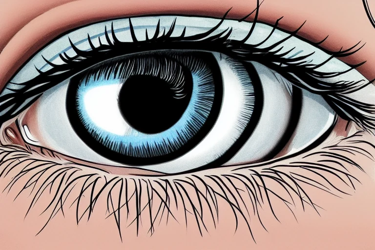
Accessory structures of the eye include the eyelids, eyelashes, eyebrows, the lacrimal apparatus, and extrinsic eye muscles. Eyelids protect the eyes during sleep, spread lubricating secretions, and control light exposure. The upper eyelid exhibits a greater range of movement than the lower eyelid as it contains the levator palpebrae superioris muscle, a muscle innervated by cranial nerve 3 in charge of lifting the eyelid. Beneath this layer is a donut-shaped muscle that encircles the eye called orbicularis oculi, which is innervated by the 7th cranial nerve, and is in charge of closing the eyelids. Twitches of the eyelids are a common and usually harmless symptom that can be caused by stress and fatigue. The space between the upper and lower eyelid that exposes the eyeball is the palpebral fissure. The lacrimal caruncle is the small, pink, globular spot at the inner corner, or the medial canthus, of the eye. It contains both oil and sweat glands. The whitish material that sometimes accumulates in that region is from these glands. [1]
Visual Pathway and Processing
Visual information travels from the retina through the optic nerve, optic chiasm, optic tract, lateral geniculate nucleus, and visual cortex. The retina contains photoreceptors that convert light into neural signals, which are processed to form images. The visual pathway maintains retinotopic organization, ensuring spatial localization. [3]
Types of Vision and Their Functions
There are various types of vision, each with specific functions. Color vision is mediated by cone photoreceptors, while night vision relies on rod photoreceptors. Motion detection is facilitated by specialized ganglion cells that respond to changes in visual scenes. [6]
Common Visual Impairments
Common visual impairments include myopia, hyperopia, astigmatism, and color blindness. Myopia and hyperopia affect the ability to focus on distant or close objects, respectively. Astigmatism distorts vision due to an irregularly shaped cornea. Color blindness impairs the ability to distinguish certain colors. [1]
Key Takeaways
- The human visual system is composed of accessory structures, photoreceptors, and neural pathways.
- Accessory structures like eyelids and lacrimal apparatus protect and lubricate the eyes.
- Visual information is processed from the retina through the optic nerve and visual cortex.
- There are different types of vision, each with specific functions.
- Common visual impairments can significantly affect daily life.
FAQ
What are the main components of the human visual system?
The main components of the human visual system include the eyes, accessory structures, photoreceptors, and a complex network of neural pathways that transmit information to the brain. [3]
How does light enter the eye and get processed to form an image?
Light enters the eye through the cornea, is focused by the lens, and is detected by photoreceptors in the retina. These photoreceptors convert light into neural signals, which are processed to form images. [3]
What are the different types of vision and how do they differ in function?
Types of vision include color vision, night vision, and motion detection. Color vision is mediated by cone photoreceptors, while night vision relies on rod photoreceptors. Motion detection is facilitated by specialized ganglion cells that respond to changes in visual scenes. [4]
What are some common visual impairments and how do they affect vision?
Common visual impairments include myopia, hyperopia, astigmatism, and color blindness. Myopia and hyperopia affect the ability to focus on distant or close objects, respectively. Astigmatism distorts vision due to an irregularly shaped cornea. Color blindness impairs the ability to distinguish certain colors. [1]
Conclusion
Vision is a complex process involving the eyes, accessory structures, and a network of neural pathways. Understanding the components and functions of the visual system can help in appreciating the marvels of human vision and the importance of maintaining eye health. [3]